- NEWS AND VIEWS
Structural studies support the idea that fibrillized tau protein, a feature of neurodegenerative disorders, acts as a prion-like template to induce misfolding in healthy tau.
By
-
Kayleigh Mason-Chalmers
-
Kayleigh Mason-Chalmers is in the Department of Chemistry and Biochemistry, the UCLA-DOE Institute for Genomics and Proteomics, and the STROBE NSF Science and Technology Center, University of California, Los Angeles (UCLA), Los Angeles, California 90095, USA.
-
-
Jose A. Rodriguez
-
Jose A. Rodriguez is in the Department of Chemistry and Biochemistry, the UCLA-DOE Institute for Genomics and Proteomics, and the STROBE NSF Science and Technology Center, University of California, Los Angeles (UCLA), Los Angeles, California 90095, USA.
-
Prions are shape-shifting, self-assembling proteins that transform from a physiologically harmless version into one that is misfolded, prone to aggregation and infectious. What makes prions and prion-like proteins capable of causing disease is their ability to act as templates, forcing normal functional proteins into a toxic form. Several proteins that are associated with neurodegenerative diseases, such as amyloid-β, α-synuclein and tau, are thought to spread in a prion-like manner1,2, but experimental support for some aspects of this hypothesis and its atomic basis have been lacking. In their paper in Nature, Lövestam et al.3 show that injecting human tau fibrils into mice causes mouse tau to take on the form of the injected structures, bolstering the theory that tau propagates similarly to prions.
doi: https://doi.org/10.1038/d41586-026-02778-w
References
Prusiner, S. B. Science 336, 1511–1513 (2012).
Article PubMed Google Scholar
Weingarten, M. D., Lockwood, A. H., Hwo, S. Y. & Kirschner, M. W. Proc. Natl Acad. Sci. USA 72, 1858–1862 (1975).
Article PubMed Google Scholar
Lövestam, S. et al. Nature https://doi.org/10.1038/s41586-026-11061-x (2026).
Article Google Scholar
Basheer, N. et al. Acta Neuropathol. Commun. 12, 52 (2024).
Article PubMed Google Scholar
Shi, Y. et al. Nature 598, 359–363 (2021).
Article PubMed Google Scholar
Fitzpatrick, A. W. P. et al. Nature 547, 185–190 (2017).
Article PubMed Google Scholar
Sawaya, M. R., Hughes, M. P., Rodriguez, J. A., Riek, R. & Eisenberg, D. S. Cell 184, 4857–4873 (2021).
Article PubMed Google Scholar
Wegmann, S., Medalsy, I. D., Mandelkow, E. & Müller, D. J. Proc. Natl Acad. Sci USA 110, 313–321 (2013).
Article Google Scholar
Scheres, S. H. W. Structure 34, 1061–1071 (2026).
Article PubMed Google Scholar
Tarutani, A. et al. FEBS Open Bio 13, 1394–1404 (2023).
Article PubMed Google Scholar
Duan, P. et al. J. Biol. Chem. 300, 107730 (2024).
Article PubMed Google Scholar
Wang, J. et al. Structure 32, 2251–2258 (2024).
Article PubMed Google Scholar
Guo, J. L. et al. J. Exp. Med. 213, 2635–2654 (2016).
Article PubMed Google Scholar
Kano, M. et al. Acta Neuropathol. Commun. https://doi.org/10.1186/s40478-026-02407-7 (2026).
Article Google Scholar
Schweighauser, M. et al. Acta Neuropathol. Commun. 11, 160 (2023).
Article PubMed Google Scholar
Rawat, P. et al. Int. J. Mol. Sci. 23, 12841 (2022).
Article PubMed Google Scholar
Burger, D. et al. Nature 648, 409–417 (2025).
Article PubMed Google Scholar
Hanger, D. P., Betts, J. C., Loviny, T. L., Blackstock, W. P. & Anderton, B. H. J. Neurochem. 71, 2465–2476 (1998).
Article PubMed Google Scholar
Watts, J. C. & Prusiner, S. B. J. Biol. Chem. 289, 19841–19849 (2014).
Article PubMed Google Scholar
Ghaemmaghami, S. et al. J. Mol. Biol. 413, 527–542 (2011).
Article PubMed Google Scholar
Download references
Competing Interests
The authors declare no competing interests.
Related Articles
-
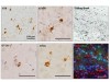 Read the paper: Prion-like transmission of human tau strains in the mouse brain
-
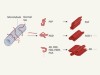 Structural strains of misfolded tau protein define different diseases
-
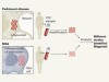 Neurodegenerative diseases distinguished through protein-structure analysis
-
See all News & Views
Subjects
Latest on:
- Structural biology
- Neurodegeneration
-
Secret watermark labels proteins as ‘made by AI’
News
-
Prion-like transmission of human tau strains in the mouse brain
Article
-
Sequence-encoded hexagonal lattices in multichannel peptide nanofibrils
Article
-
Landmark map of human brain’s gene activity holds clues to Alzheimer’s disease and more
News
-
Vast cellular gene-expression atlas could transform how scientists understand brain ageing and disease
News & Views
-
Single-cell atlas of transcriptomic vulnerability across brain disorders
Article